CEPHASONICS ULTRASOUND
  • Home
  • OEM ultrasound
  • Ultrasound Systems
  • Software
  • Quantitative Ultrastround
  • AI and Ultrasound
  • Cephasonics Products
  • Custom engineering and manufacturing
  • News
  • Contact Us
  • Accessories and Interfaces
  • Remote control
  • Home
  • OEM ultrasound
  • Ultrasound Systems
  • Software
  • Quantitative Ultrastround
  • AI and Ultrasound
  • Cephasonics Products
  • Custom engineering and manufacturing
  • News
  • Contact Us
  • Accessories and Interfaces
  • Remote control
Search by typing & pressing enter

YOUR CART

Cephasonics Dual-Mode Elastography

Overview

Cephasonics Ultrasound Elastography systems provide the ability to execute a variety of ultrasound push functions as well as full-function ultrasound imaging.  This combination of capabilities makes it perfect for research projects into ultrasound elastography as well as development of commercial diagnostic elastography applications.

Picture
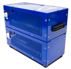
The Cicada PX Elastography systems come in 128-256 channel configurations and includes an Ubuntu version of a Linux-based server configured with one or more GPUs and PCIe interfaces to the ultrasound system. 


Dual-Mode Elastography

The Cephasonics Elastography system can be configured in 2 ways.  In a single system configuration, a single ultrasound transducer is used to both generate acoustic radiation force in tissue and then image the tissue displacement response.  

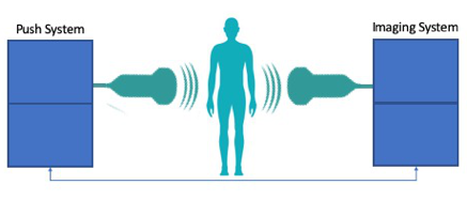
In a Dual-Mode configuration, a Cicada PX push system is paired with a second Cicada imaging system.

This enables the simultaneous push/imaging with 2 probes – one connected to each of the systems.  In this configuration, the Cicada PX system is connected to and synchronized with the second Cicada imaging system. 

This allows you to push with one probe and at the same time image using a second probe on the other system. 

Picture

Elastography Specifications
Supported push modes:
  • Beam forming imaging
  • Ultrafast Imaging
  • Shear Wave Elastography

Capabilities
  • Push Burst can generate 4 pulses per second or up to 50,000 cycles per second with a single pulse (Constant droop depends on probe impedance and frequency)
  • 100 volts peak to peak
  • Collect up to 100 frames per push
  • Uses the “Loupas” algorithm to measure phase difference between frames.
  • Allows direct access to data to do measurements and generate motion data
  • Track and collect up to 32KHz PRF with interleaved mode and up to 10KHz PRF for non-interleaved mode

Note: Muxing is not supported on Push systems
Probes supported for push pulse
  • Cephasonics 7Mhz linear probe
  • Cephasonics 3MHz curved probe
  • Custom push probes utilizing a Cephasonics Back shell adapter kit

Elastography Software Features:
  • The system provides 4 separate modules
    • Push – create a push pulse with the system
    • Data collection – collect imaging data after the push
    • Reconstruct – create images from the imaging data
    • Compute – calculate the phase differences (Loupas algorithm) and other functions
  • The software provides a script-based control interface for operating the push and imaging functions of the system.  The run-time script allows you to enter specific parameters into a file and execute it for push/image sequences


Imaging Specifications
Supported imaging modes:
  • BMode, Harmonic Imaging
    • Simplex
    • Image Enhancement
    • Spatial compounding
    • Multi focal zone
    • Standard 2D Imaging controls
    • Advanced 2D Imaging controls
  • CFM
    • Duplex
    • Standard CFM controls
    • Advanced CFM controls
Imaging Capabilities:
  • Fine-grain control of transmit and receive parameters for channel capture and beamforming modes of operations
    • Transmit wave definition
    • Transmit/Receive delays and weights
    • Probe mux configuration per firing
  • User-configurable input and output triggering to synchronize ultrasound firing with external systems
  • Fully programmable AFE and TGC parameters
  • Fully programmable depth-based frequency tracking demodulation and decimation filtering (TFC)
  • Example programs illustrate key advanced features
  • B-mode, harmonic imaging, Doppler modes, including CFM, PDI and PWD, and M-mode
  • Data rates to 3.6Gbps over PCI-express v1.1 (x4)
  • Frame rates of up to 28fps over USB 2.0 interface at 20cm depth
  • Resolution: lateral 0.5mm, axial 0.5mm
  • Geometrical precision:  0.1mm or 1 pixel
  • Spatial compounding
  • Multi focal zone (max 4 zones) , multi frequency
  • Scan angle from 0 deg to 150 deg, at 1 deg increment
  • Image depth: 1mm to 30cm
Imaging Transmit
  • 256 pulse shape entries, each 248 pulses at 160MHz
  • 8192 sample long pulses for “chirp” signal
  • Output pulse rate: 100Hz to 40MHz
  • Pulse repetition counter 1...32
  • Pulse duty cycle adjustment to 6.125ns precision
  • Arbitrary wave form definition
  • Transmit aperture definition
    • Arbitrary channel enabling
    • Apodization F#:  1/16...16
  • Delay resolution: 6.125ns
  • 3-level or 5-level pulser configurations available
  • Output jitter (at 5MHz): 25ps
  • Phase Noise (at 5MHz, 1KHz offset): −116 dBc/Hz
Receive
  • ADC: 12-bit 68.5dBFS, 10.5ENOB
  • Ultra-low 2.4dB noise figure
  • Input frequency range: 1MHz to 18MHz
  • Sampling rate: 20MHz and 40MHz
  • Close-in phase-noise (at 1KHz offset from 3.5MHz carrier): -140dBc/Hz
  • Channel Capture
    • 8K samples per channel buffer
    • Programmable high pass filter
    • Programmable decimation filter
Beamforming
  • Continuous receive focus and apodization: delay and weight coefficients updated on every sample
    • 1-beam and 2-beam parallel processing
    • Preset window functions
    • Programmable Time Frequency Control in 32 zones
 

Cephasonics Ultrasound is a division / registered dba of White Eagle Sonic Technologies, Inc.
​

Products

Systems
Accessories
Software
Consulting
​Probes

Company

About the company

Support

Contact
News
FAQ
Terms of Use

Picture
© COPYRIGHT 2025.
White Eagle Sonic Technologies. Inc.
ALL RIGHTS RESERVED.